RABBIT ANTI MOUSE MCP-1 (AAM43) 製品に関するお問い合わせは、こちらのフォームをご利用ください。
| 販売元 | |
|---|---|
| 製品タイプ | Antibody - Polyclonal |
| 画像データ |
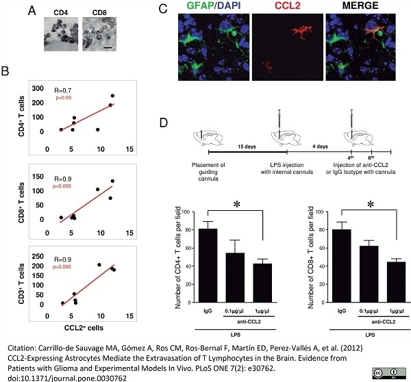 Rabbit anti Mouse MCP-1 antibody used for the identification of MCP-1 expressing cells in murine glioma by indirect immunofluorescence on formailin fixed vibratome sections. Image caption: CCL2+ cells are GFAP+ astrocytes in human glioma. (A) Confocal images show co-localization of CCL2-expressing cells (red) with GFAP (green) in glioma. Insert shows a detail of a single CCL2-expressing cell (red) co-localizing with GFAP (green) and counterstained with DAPI (blue). Scale bar: 30 μm. (B) Areas of infiltration of CD3+ T-cells (green) coincide with the areas of CCL2-expressing cells (red) in glioma. Confocal images show a tumorigenic area, infiltrated with CD3+ T-cells (green) in an area with numerous CCL2-expressing cells (red). The immunostaining was combined with a counterstaining with DAPI to detect the nucleus (blue). Scale bar: 50 μm. (C) Examples of characteristic CD3 infiltration in samples of glioma. CD3+ T-cells can be observed grouped in BVs (BV, limited by broken red line in 1 and 3) but also infiltrated in the parenchyma (2, 4, blue insert magnified in 5). (D) CCL2 expression correlates with the infiltration of T-cells in the tumor areas. The quantification in serial sections of the number of T-cells, either infiltrated or located in the BV lumen, revealed that the level of infiltration of CD3+ T-cells in the parenchyma (green) is positively correlated with the level of CCL2 expression (BV; BV lumen). From: Citation: Carrillo-de Sauvage MA, Gómez A, Ros CM, Ros-Bernal F, Martín ED, Perez-Vallés A, et al. (2012) CCL2-Expressing Astrocytes Mediate the Extravasation of T Lymphocytes in the Brain. Evidence from Patients with Glioma and Experimental Models In Vivo. PLoS ONE 7(2): e30762. doi:10.1371/journal.pone.0030762. ウサギ抗マウスMCP-1抗体は、CCL2、小誘導性サイトカインA2、または血小板由来成長因子誘導性タンパク質JEとしても知られるマウス単球走化性タンパク質-1(MCP-1)を認識します。 マウスMCP-1は148アミノ酸〜16kDaの分泌された単球走化性因子です ケモカインMCP-1は、単球、血管内皮細胞、平滑筋細胞、糸球体メサンギウム細胞、骨芽細胞などのさまざまな細胞によって発現されます。 MCP-1は、血中単球および組織マクロファージの炎症反応において重要な役割を果たします(Conti and DiGioacchino2001)。 |
| 価格 | ¥105,000 |
| カタログ番号 |
AAM43
|
| 容量 | 0.1 mg |
| 用途 | Functional Assay, Immunofluorescence, Western Blotting, ELISA |
| フォーマット / 標識 | Purified |
| 交差性 | Mouse |
| クローン | |
| 免疫動物 | Rabbit |
| データシート | |
| 詳細情報 | 製品詳細情報はこちらをクリック(論文情報、その他の画像データなど) |
※弊社製品は、最寄りの販売代理店を通してご注文ください。ご不明な場合、弊社までご連絡ください。
※製品および技術的なご質問、資料のご請求などございましたらこちらまでご連絡ください。
※まとめ買い、大量購入、カスタマイズサービスについては、販売代理店へご確認ください。
※表示価格はメーカー希望小売価格(税別)です。詳細は販売代理店へご確認ください。なお、製品情報、価格等は予告なく変更される場合がございます。
※用途に[*]がついているものは一定の条件下での使用が推奨されたものです。製品詳細ページよりデータシートにてご確認ください。
バイオ・ラッド ラボラトリーズ株式会社
住所:〒140-0002 東京都品川区東品川2-2-24 天王洲セントラルタワー20F
電話:03-6361-7000
FAX:03-5463-8480