SHEEP ANTI HUMAN VON WILLEBRAND FACTOR (AHP062T) 製品に関するお問い合わせは、こちらのフォームをご利用ください。
| 販売元 | |
|---|---|
| 製品タイプ | Antibody - Polyclonal |
| 画像データ |
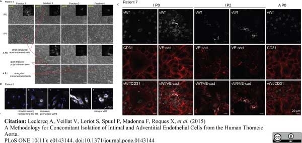 Sheep anti Human von Willebrand Factor antibody used for the evaluation of von Willebrand factor expression in isolated aortic cell preparations by immunofluorescence. Image caption: Characterization of the isolated cells. (A) Representative phase contrast micrographs show the cell population collected at each of the four consecutive rounds of digestion (well 1 to 4) on the intimal (I) and adventitial (A) side of the aorta. In both cases, three main phenotypes were observed: small polygonal cells forming areas of cobblestone appearance, giant mono- or multinucleated cells and elongated cells. All three phenotypes were also detected at P1. Immunofluorescence analysis performed on each P0 population identified ECs by positive vWf staining (insert). The small polygonal mononucleated cells and the giant mono- or polynucleated morphologies, stained both for vWf. Rare elongated cells could also be vWf positive, their unusual shape likely being due to surrounding peer pressure. Magnification of the phase contrast pictures and immunofluorescence images is 10x and 63x, respectively. (B) Immunofluorescent micrographs showing the diversity of vWf presentation. vWf staining performed on adventitial aortic endothelial cells shows diverse and typical vWF staining patterns such as that of elongated WPBs throughout the cell body (d and e), residual vWf granules after degranulation of WPB or seen in cross section (a), reticular vWf around the nucleus corresponding to vWf re-synthesis after degranulation (b and c) or extracellular string of vWf (e). (C) Immunofluorescent micrographs of isolated cells double-stained for vWf together with either CD31 or VE-cadherin validates vWf staining. Small polygonal mono-nucleated cells and most of the giant mono- or polynucleated vWf positive cells also stained for CD31 or VE-cadherin at P0 and P2 and in IEC and AEC. From: Leclercq A, Veillat V, Loriot S, Spuul P, Madonna F, Roques X, et al. (2015) A Methodology for Concomitant Isolation of Intimal and Adventitial Endothelial Cells from the Human Thoracic Aorta. PLoS ONE 10(11): e0143144. This image is from an open access article distributed under the terms of the Creative Commons Attribution License. ヒツジ抗ヒトフォンウィルブランド因子抗体は、内皮細胞および巨核球で合成され、血液中を循環する糖タンパク質であるヒトフォンウィルブランド因子を、第VIII因子との結合により非共有複合体として認識します。 |
| 価格 | 販売終了 |
| カタログ番号 | AHP062T |
| 容量 | 0.1 ml |
| 用途 | Flow Cytometry, Immunohistology - Frozen Sections*, Immunofluorescence, ELISA |
| フォーマット / 標識 | Purified |
| 交差性 | Human, Rat, Pig |
| クローン | |
| 免疫動物 | Sheep |
| データシート | |
| 詳細情報 | 製品詳細情報はこちらをクリック(論文情報、その他の画像データなど) |
※弊社製品は、最寄りの販売代理店を通してご注文ください。ご不明な場合、弊社までご連絡ください。
※製品および技術的なご質問、資料のご請求などございましたらこちらまでご連絡ください。
※まとめ買い、大量購入、カスタマイズサービスについては、販売代理店へご確認ください。
※表示価格はメーカー希望小売価格(税別)です。詳細は販売代理店へご確認ください。なお、製品情報、価格等は予告なく変更される場合がございます。
※用途に[*]がついているものは一定の条件下での使用が推奨されたものです。製品詳細ページよりデータシートにてご確認ください。
バイオ・ラッド ラボラトリーズ株式会社
住所:〒140-0002 東京都品川区東品川2-2-24 天王洲セントラルタワー20F
電話:03-6361-7000
FAX:03-5463-8480