RABBIT ANTI HUMAN TGN46 (AHP1586) 製品に関するお問い合わせは、こちらのフォームをご利用ください。
| 販売元 | |
|---|---|
| 製品タイプ | Antibody - Polyclonal |
| 画像データ |
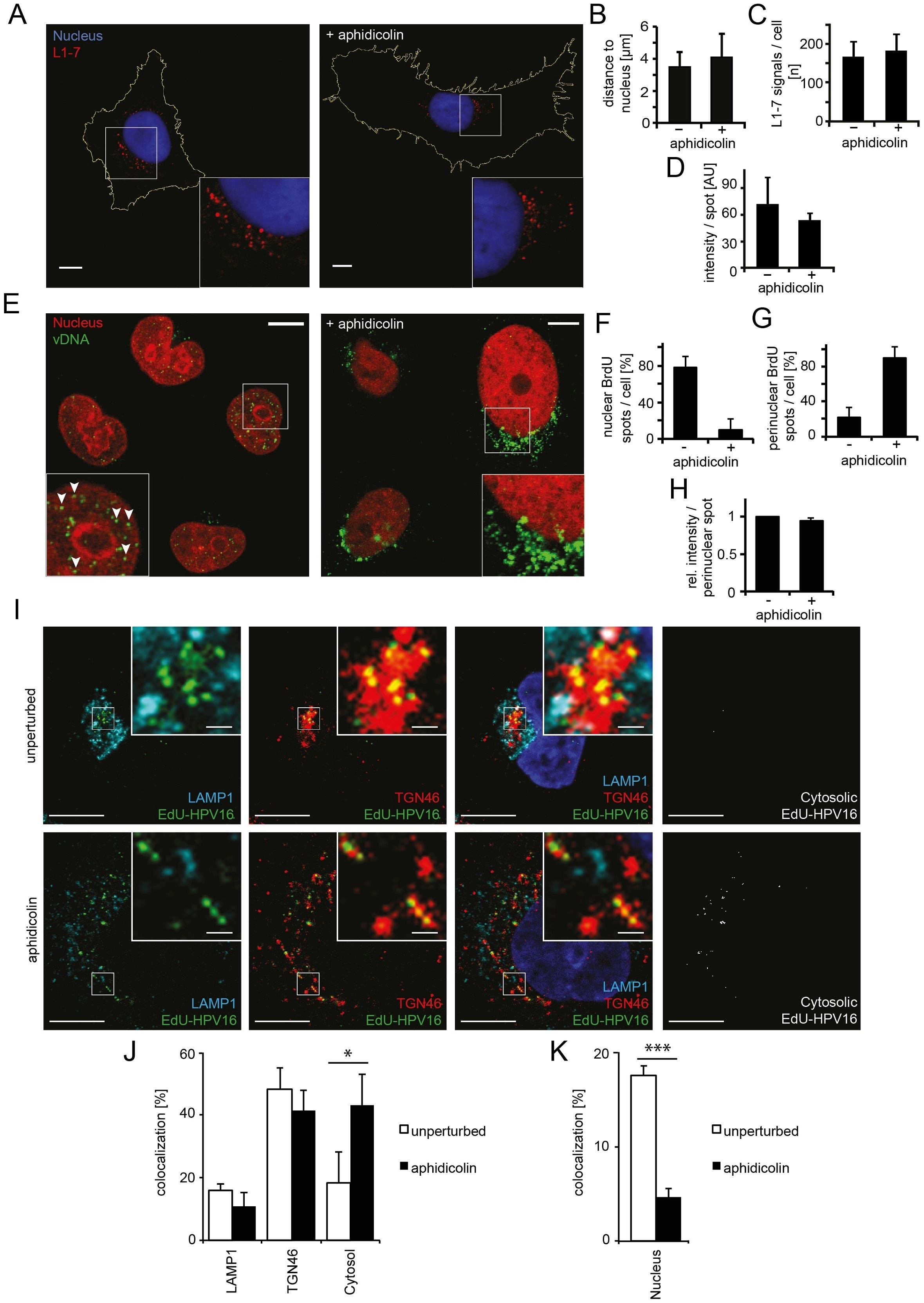 Rabbit anti Human TGN46 antibody used for the localisation of the trans golgi network in HeLa cells by immunofluorescence. Image caption: Nuclear import of HPV16 vDNA is blocked in interphase cells. (A) Aphidicolin-treated or untreated HeLa cells were infected with HPV16-GFP. At 16 h p.i., cells were fixed and immunostained with the L1-7 antibody detecting endosomal, conformationally altered virions. Depicted are confocal sections of untreated (left) or aphidicolin-treated (right) cells. (B) As in (A) with quantification of the three-dimensional distance of L1-7 signals to the nuclear border (in ツオm) ツア SD. (C) As in (A) with quantification of the number of discernible L1-7 spots/cell. (D) As in (A) with quantification of the fluorescent intensity (in arbitrary units, AU) of individual L1-7 spots. (E) HeLa H2B-mCherry cells were infected with BrdU-HPV16. Cells were immunostained for BrdU to detect the vDNA after fixation at 24 h p.i.. Depicted are confocal sections of aphidicolin-treated (interphase, right) or untreated (left) cells. Mostly, the vDNA localized intranuclearly as discrete spots in untreated cells (arrowheads) or exclusively perinuclearly in aphidicolin-treated cells. (F) As in (E) with quantification of intranuclear BrdU spots/cell. The number of intranuclear spots is given relative to the total number of cellular spots. (G) As in (E) with quantification of perinuclear BrdU spot/cell as in (F). (H) As in (E) with quantification of the signal intensities of individual perinuclear BrdU spots relative to untreated infected cells. (I) HeLa cells were treated with or without aphidicolin for 16 h prior to infection. 20 h after infection with EdU-HPV16 (green), cells were fixed and immunostained for TGN46 (red) and LAMP1 (light blue). The nucleus was stained by Hoechst (dark blue). Depicted are single confocal sections. The cytosolic EdU-HPV16 signal is depicted after substraction of the EdU-HPV16 signals colocalizing with LAMP1, TGN46 and the nucleus. (J) As (I) with quantification of signal colocalization of EdU-HPV16 with LAMP1 or TGN46. Cytosolic EdU-HPV16 amounts were defined as signals that did not colocalize with LAMP1, TGN46, or the nucleus (see below). (K) As in (I) with quantification of signal colocalization of EdU-HPV16 with the nuclear stain (Hoechst). Statistical significance was determined by a two-tailed, independent t-test; P-values: * <0.05; *** <0.001. All scale bars: 10 ツオm. From: Aydin I, Weber S, Snijder B, Samperio Ventayol P, Kテシhbacher A, Becker M, et al. (2014) Large Scale RNAi Reveals the Requirement of Nuclear Envelope Breakdown for Nuclear Import of Human Papillomaviruses. PLoS Pathog 10(5): e1004162. ウサギ抗ヒトTGN46抗体は、TGN46、TGN38ホモログまたはトランスゴルジネットワークタンパク質TGN51としても知られるトランスゴルジネットワーク内在性膜タンパク質2を認識します。 TGN46は、トランスゴルジネットワーク膜に関連する480アミノ酸〜110kDaのシングルパスI型膜貫通糖タンパク質です。 TGN46は、ヒトのトランスゴルジネットワークで利用可能な最良のマーカーであると報告されています。 |
| 価格 | ¥109,000 |
| カタログ番号 |
AHP1586
|
| 容量 | 0.1 ml |
| 用途 | Western Blotting, Immunofluorescence* |
| フォーマット / 標識 | Serum |
| 交差性 | Human, Primate |
| クローン | |
| 免疫動物 | Rabbit |
| データシート | |
| 詳細情報 | 製品詳細情報はこちらをクリック(論文情報、その他の画像データなど) |
※弊社製品は、最寄りの販売代理店を通してご注文ください。ご不明な場合、弊社までご連絡ください。
※製品および技術的なご質問、資料のご請求などございましたらこちらまでご連絡ください。
※まとめ買い、大量購入、カスタマイズサービスについては、販売代理店へご確認ください。
※表示価格はメーカー希望小売価格(税別)です。詳細は販売代理店へご確認ください。なお、製品情報、価格等は予告なく変更される場合がございます。
※用途に[*]がついているものは一定の条件下での使用が推奨されたものです。製品詳細ページよりデータシートにてご確認ください。
バイオ・ラッド ラボラトリーズ株式会社
住所:〒140-0002 東京都品川区東品川2-2-24 天王洲セントラルタワー20F
電話:03-6361-7000
FAX:03-5463-8480