RAT ANTI MOUSE CD169 (MCA947) 製品に関するお問い合わせは、こちらのフォームをご利用ください。
| 販売元 | |
|---|---|
| 製品タイプ | Antibody - Monoclonal |
| 画像データ |
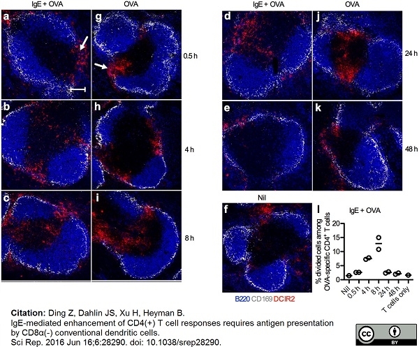 AlexaFluor®647 conjugated Rat anti Mouse CD169 antibody, clone MOMA-1 used to demonstrate marginal zone metallophils by immunofluorescence. Image caption: DCIR2+ cDCs migrate from the marginal zone bridging channel to the T cell zone after immunization. Spleens from BALB/c mice (n窶・窶・ per time point) immunized with 250窶・mu;g IgE anti-OVA pre-mixed with 100窶・mu;g OVA or with 100窶・mu;g OVA alone were harvested after 0.5, 4, 8, 24, or 48窶栄. One unimmunized mouse (Nil) was used as control. (a窶徒) Half of each spleen was snap-frozen and non-consecutive spleen sections were stained and analyzed by confocal microscopy. Localization of DCIR2+ cDCs in spleens harvested at indicated time points after immunization was followed. B220+ B cells, blue; CD169+ metallophilic macrophages, grey; DCIR2+ cDCs, red. Marginal zone bridging channels are indicated with arrows in (a,g). Images show representative areas (640窶・mu;m窶嘉冷・40窶・mu;m) of 3窶・窶欝 cell zones from 2 non-consecutive sections of each sample in every group. Scale bar represents 100窶・mu;m. Data represent one experiment where mice were immunized with IgE-OVA or OVA alone and one where they were immunized with IgE-OVA. (l) The other halves of the spleens from mice immunized with IgE-OVA complexes in (a窶兎) were prepared into single cell suspensions and 6窶嘉冷・05 cells were used as APCs in co-cultures with 105 CFSE-labeled CD4+ T cells isolated from DO11.10 splenocytes. Percentages of divided cells among OVA-specific CD4+ T cells after incubation for 3 days with APCs taken from an unimmunized mouse (Nil) or from mice immunized with IgE-OVA complexes are quantified by flow cytometry as shown in Fig. 3. CD4+ T cells cultured alone were used as negative control. Each circle represents one mouse and the lines represent the mean values. From: Ding Z, Dahlin JS, Xu H, Heyman B. IgE-mediated enhancement of CD4(+) T cell responses requires antigen presentation by CD8α(-) conventional dendritic cells. Sci Rep. 2016 Jun 16;6:28290. ラット抗マウスCD169、クローンMOMA-1は、sialoadhesinまたはSiglec-1としても知られるマウスCD169を認識します。 CD169は、脾臓の辺縁帯の金属親和性物質、リンパ節の被膜下マクロファージ、骨髄の間質マクロファージなど、特定のマクロファージ集団によって発現されるレクチン様受容体です(Morris et al.1991)。 CD169は、17の免疫グロブリン様ドメインを含む約185 kDaのシアル酸結合受容体です(Crocker et al.1992)。 CD169の発現は、培養中のマクロファージで血清因子によって誘導され、サイトカイン曝露によってさらに調節されます(McWilliam et al.1992)。 ラット抗マウスCD169、クローンMOMA-1は、特定のマクロファージ集団のin vivoでの枯渇に使用されています(Kraal et al.1988)。 |
| 価格 | 販売終了 |
| カタログ番号 | MCA947 |
| 容量 | 2 ml |
| 用途 | Snap Frozen, Acetone Fixed Immunohistological Sections, Immunofluorescence |
| フォーマット / 標識 | S/N |
| 交差性 | Mouse |
| クローン | MOMA-1 |
| 免疫動物 | Rat |
| データシート | |
| 詳細情報 | 製品詳細情報はこちらをクリック(論文情報、その他の画像データなど) |
※弊社製品は、最寄りの販売代理店を通してご注文ください。ご不明な場合、弊社までご連絡ください。
※製品および技術的なご質問、資料のご請求などございましたらこちらまでご連絡ください。
※まとめ買い、大量購入、カスタマイズサービスについては、販売代理店へご確認ください。
※表示価格はメーカー希望小売価格(税別)です。詳細は販売代理店へご確認ください。なお、製品情報、価格等は予告なく変更される場合がございます。
※用途に[*]がついているものは一定の条件下での使用が推奨されたものです。製品詳細ページよりデータシートにてご確認ください。
バイオ・ラッド ラボラトリーズ株式会社
住所:〒140-0002 東京都品川区東品川2-2-24 天王洲セントラルタワー20F
電話:03-6361-7000
FAX:03-5463-8480